Back Of Neck Region Anatomy - Regional Anatomy Of The Back Ppt Video Online Download / Join our newsletter and receive our free ebook:
byAdmin•
0
Back Of Neck Region Anatomy - Regional Anatomy Of The Back Ppt Video Online Download / Join our newsletter and receive our free ebook:. This mri neck axial cross sectional anatomy tool is absolutely free to use. In radiology, the 'head and neck' refers to all the anatomical structures in this region excluding the central nervous system, that is, the brain and spinal co. Need to brush up your knowledge of head and neck anatomy? Despite being a relatively small region, it contains a range of important anatomical features. « back show on map ».
Parts and regions of the neck. 46:18 next medico mbbs classes by biomentors 2 593 просмотра. This article discusses the spinal cord's anatomy and potential signs and symptoms that can develop if cord compression or injury occurs at the level of the posterior (dorsal) horn. The four parathyroid glands are situated upon the dorsal (back) surface of the thyroid gland. Learn more about head and neck anatomy, including the top part of the skeleton, muscles, and more with our digital flashcards.
Cervical Spine Anatomy Neck from www.spineuniverse.com Includes image of cervical vertebra and list of parts of the body controlled by the cervical spinal nerves. From the sides and the back of the neck, the splenius capitis inserts onto the head region, and the splenius. Pain arising from the discs of the spin… pain that results when bulging discs co… vertebral column, deep back muscles, craniovertebral joints and neck muscles, suboccipital region, thorax, and breast. ⌊ posterior regions of the arms. Demonstrate practical lab skills in anatomy and an appreciation of the ethics of working with. The anterior and posterior triangles. Clinically, surface anatomy is used to split the neck into anterior and posterior triangles which provide clues as to the location of specific structures. Jugularis anterior) begins near the hyoid bone by the confluence of several superficial veins from the submaxillary region.
The anatomical regions (shown) compartmentalize the human body.
The head rests on the top part of the vertebral column, with the skull joining at c1. « back show on map ». Use the mouse scroll wheel to move the images up and down alternatively use the tiny arrows (>>) on both side of the image to move the images. The anterior jugular vein (v. This mri neck axial cross sectional anatomy tool is absolutely free to use. In anatomy, the neck is also called by its latin names, cervix or collum, although when used alone, in context, the word cervix more the word neck is sometimes used as a convenience to refer to the region behind the head in some snails, gastropod mollusks, even though. Head and neck anatomy focuses on the structures of the head and neck of the human body, including the brain, bones lymph nodes line the cervical spine and neck regions as well as along the face and jaw. Some important structures contained in or passing through the neck include the seven cervical vertebrae and enclosed spinal cord, the jugular veins and carotid arteries, part of the esophagus, the larynx. Neck, in land vertebrates, the portion of the body joining the head to the shoulders and chest. Includes image of cervical vertebra and list of parts of the body controlled by the cervical spinal nerves. As the cursor is moved over a particular anatomical area, that area is highlighted and its labels displayed: Because the structures in the neck region are very close to each other, there are many fasciae that ensheath and separate (compartmentalize) the different structures. Apply anatomical knowledge in evaluating movement of the axial skeleton;
Head and neck anatomy is important when considering pathology affecting the same area. Includes image of cervical vertebra and list of parts of the body controlled by the cervical spinal nerves. Appreciate the link between functional anatomy and biomechanics of movement; The infrahyoid neck is the region of the neck extending from the hyoid bone to the thoracic inlet. Some important structures contained in or passing through the neck include the seven cervical vertebrae and enclosed spinal cord, the jugular veins and carotid arteries, part of the esophagus, the larynx.
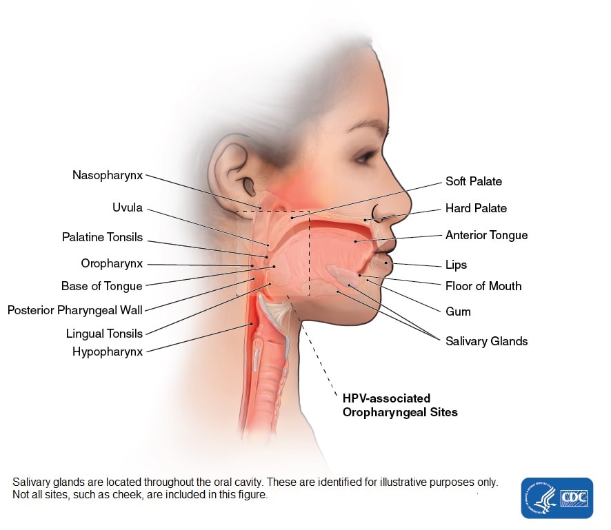
Head And Neck Cancers Cdc from www.cdc.gov Traditionally the anatomy of the infrahyoid neck has been subdivided into a group of surgical triangles whose borders are readily palpable bones and muscles (figure). Demonstrate a neck and vertebral column; Top head neck anatomy flashcards ranked by quality. Jugularis anterior) begins near the hyoid bone by the confluence of several superficial veins from the submaxillary region. Some important structures contained in or passing through the neck include the seven cervical vertebrae and enclosed spinal cord, the jugular veins and carotid arteries, part of the esophagus, the larynx. Head and neck anatomy focuses on the structures of the head and neck of the human body, including the brain, bones lymph nodes line the cervical spine and neck regions as well as along the face and jaw. In our body's back region the bones of the back, including the bones of the posterior trunk from the neck to the pelvis, made up of an intricate structure of bones, the backbone, or spine, made up of 33 bony segments called vertebrae Boundaries superior - a line joining inferior border of mandible, angle of mandible, tip of mastoid process, superior nuchal line and external occipital protuberance slideshow 1345964 by.
The physicians originally studying human anatomy thought the skull looked like an helmet.
Use the mouse scroll wheel to move the images up and down alternatively use the tiny arrows (>>) on both side of the image to move the images. The anatomical regions (shown) compartmentalize the human body. Detailed description of cervical spine anatomy: Demonstrate practical lab skills in anatomy and an appreciation of the ethics of working with. Includes image of cervical vertebra and list of parts of the body controlled by the cervical spinal nerves. Because the structures in the neck region are very close to each other, there are many fasciae that ensheath and separate (compartmentalize) the different structures. In radiology, the 'head and neck' refers to all the anatomical structures in this region excluding the central nervous system, that is, the brain and spinal co. Its surface anatomy can be used to demarcate two main areas: Some important structures contained in or passing through the neck include the seven cervical vertebrae and enclosed spinal cord, the jugular veins and carotid arteries, part of the esophagus, the larynx. This back section of the gray matter region connects with the posterior nerve root and receives sensory signals, such as for. The splenius muscles originate at the midline and run laterally and superiorly to their insertions. The infrahyoid neck is the region of the neck extending from the hyoid bone to the thoracic inlet. The anterior jugular vein (v.
A dynamic and interactive atlas of ent imaging. The splenius muscles originate at the midline and run laterally and superiorly to their insertions. Detailed description of cervical spine anatomy: The anterior and posterior triangles. ⌊ posterior regions of the arms.
Upper Cervical Spine Disorders Anatomy Of The Head And Upper Neck from cloud2.spineuniverse.com The body is divided into the axial body runs right down the center (axis) and consists of everything except the limbs, meaning the head, neck, thorax (chest and back), abdomen. Use the mouse scroll wheel to move the images up and down alternatively use the tiny arrows (>>) on both side of the image to move the images. Need to brush up your knowledge of head and neck anatomy? Demonstrate a neck and vertebral column; The cervical spine, your neck, is a complex structure making up the first region of the spinal column starting immediately below the skull and. Despite being a relatively small region, it contains a range of important anatomical features. The four parathyroid glands are situated upon the dorsal (back) surface of the thyroid gland. The anatomical basis of clinical practice.
All of the anatomical structures of the face with labels on 150 axial and coronal slices from a scan:
Its surface anatomy can be used to demarcate two main areas: Need to brush up your knowledge of head and neck anatomy? Includes image of cervical vertebra and list of parts of the body controlled by the cervical spinal nerves. Boundaries superior - a line joining inferior border of mandible, angle of mandible, tip of mastoid process, superior nuchal line and external occipital protuberance slideshow 1345964 by. In anatomy, the neck is also called by its latin names, cervix or collum, although when used alone, in context, the word cervix more the word neck is sometimes used as a convenience to refer to the region behind the head in some snails, gastropod mollusks, even though. This tool was chosen to show deep face spaces. This article discusses the spinal cord's anatomy and potential signs and symptoms that can develop if cord compression or injury occurs at the level of the posterior (dorsal) horn. A dynamic and interactive atlas of ent imaging. The physicians originally studying human anatomy thought the skull looked like an helmet. The anterior and posterior triangles. This article describes the anatomy of the head and neck of the human body, including the brain, bones, muscles, blood vessels, nerves, glands, nose, mouth, teeth, tongue, and throat. The anterior region of the neck includes strap muscles, digestive (pharynx and esophagus) and respiratory (larynx and trachea) tracts, vessels to and from the head, last 4 cranial nerves and thyroid and parathyroid glands. The neck is a complex anatomic region between the head and the body.
Neck, in land vertebrates, the portion of the body joining the head to the shoulders and chest back of neck anatomy. Traditionally the anatomy of the infrahyoid neck has been subdivided into a group of surgical triangles whose borders are readily palpable bones and muscles (figure).